Hol lab

Glia biology of brain diseases

The aim of our research is to understand the role of glia in various brain diseases, such as Alzheimer’s disease and glioma, and to study the regenerative potential of the astrocytic stem cells in brain diseases.

Glial cells consist of astrocytes, microglia and oligodendrocytes, and are the most predominant cell type in the brain. Glia regulate local microcirculation, modulate the communication between neurons, and are the stem cells in the brain. Over the years, it has become increasingly clear that glia are involved in many brain diseases. Astrocytes can become reactive, which is likely to affect brain functioning. We use various techniques and model systems to unravel the molecular and functional changes in glia and how this contributes to the pathology of various brain diseases. The ultimate goal of our research is to contribute to developing therapeutic strategies to control reactive gliosis and to stimulate the endogenous repair capacity of the astrocytic stem cells in brain diseases.

Neuron-glia interactions and cognitive decline
Astrocytes (GFAP, green), amyloid beta plaques (red) and nuclei (blue) in a 9 month old Alzheimer mouse model (APP/PS1).
Alzheimer’s disease (AD) is a devastating neurodegenerative disease that is the biggest cause of dementia. It is characterized by neurofibrillary tangels, amyloid β (Aβ) plaques and reactive gliosis, which leads to neuronal degeneration and cognitive decline. Glial cells are essential in regulating optimal neuronal communication and synaptic transmission. Astrocytes become reactive in AD, changing their gene expression and function. We hypothesize that astrocytes lose their normal role in supporting neuronal function when they get in contact with Aβ plaques, resulting in loss of synaptic connections and neuronal death. With the APP-PS1 mouse model we study the effect of attenuated astrogliosis on memory during AD and aim to identify genes involved when the astrocytes become reactive. We also study the events that take place in the early stages of AD, where oligomeric forms of Aß protein may already negatively affect synapses leading to the initial cognitive impairments. A combination of techniques, including slice electrophysiology and calcium imaging, are used to reveal functional changes at the microcircuit level and which underlying mechanisms are involved in the APP-PS1 mouse model. We aim to elucidate changes in synaptic transmission, as well as whether pharmacological intervention prevents these changes from happening.

Alzheimer’s disease pathology in the human hippocampus: neuronal Tau-protein (green), reactive astrocytes (GFAP, red) and nuclei (blue).
Cognitive decline can also be the result of a stroke. Aneurysmal subarachnoid hemorrhage (SAH) is a bleeding into the subarachnoid space caused by a rupture of an intracranial arterial aneurysm. Aneurysmal SAH represents 5% of all stroke types and affects about 9 per 100,000 individuals annually. SAH occurs at a young age; half of the patients are younger than 50. The prognosis is poor as about a third of the patients will die within the first weeks, and another third will remain permanently disabled. We are studying the role of Subarachnoid Hemorrhage (SAH)-induced gliosis on cognitive impairment and a potential new therapeutic approach to promote recovery of brain function after SAH.
The two faces of glial stem cells: regeneration and glioma
Neural progenitors in the astrocytic ribbon of the adult human brain.
The subventricular zone (SVZ) is a principal source of neural progenitors in the adult mammalian brain. In the rodent brain this region continuously generates new neurons for the olfactory bulb. The astrocytes in the SVZ have been identified as multipotent neural progenitors, which are the precursors of newly generated neurons in the adult brain. Recently it has also been shown that human astrocytes from the SVZ area, that form a ribbon lining the ventricles, divide in vivo and produce multipotent self-renewing neurospheres in vitro. Our group has found a specific marker for the human astrocytic ribbon. We have shown that astrocytes in the human brain germinal zones express a specific isoform of the glial fibrillary acidic protein (GFAP), i.e. GFAP-δ. Currently we are looking for novel targets to stimulate brain repair in Parkinson’s disease.

Perivascular invading glioma cells within ex vivo brain slices
Overproliferation of neural stem cells can result in glioma. Gliomas are the most common form of malignant primary brain tumours in adults. A treatment remains to be found, which is mostly due to the highly invasive nature of these type of tumours. It is therefore important to decipher the mechanism of the cellular behaviour of glioma tumours. We study how the intermediate filament (IF) network, specifically the IF protein glial fibrillary acidic protein (GFAP), is involved in the cellular behaviour of glioma cells. Using a 3D model, we study the GFAP isoforms and how they influence the proliferative growth and invasiveness of patient-derived glioblastoma cells in organotypic brain slices.

Perivascular invading glioma cells within ex vivo brain slices
It has also recently become clear that the protein IkappaBzetta plays an important role in the development of the most severe type of glioma, glioblastoma (GBM). Since patients with this type of brain tumor often don’t have a good prognosis, it is important to clarify the underlying mechanism. We aim to understand the molecular mechanism of the IkappaBzetta pathway in patient-derived GBM cell lines. We also intend to unravel if certain modulators can be controlled in a targeted manner, which could be used as novel targets for possible anti-tumour treatments.

Another focus is the interaction between GBM and its micro-environment. We make use of 2D co-culture models using primary patient tumor cells and tumor-derived immune cells, and a 3D patient-derived glioblastoma invasion model using cerebral organoids. By promoting the anti-tumour resonse of immune cells through overexpression of Class II Transactivator, the master regulator of MHC class II, we aim to better understand the role of the immune system in GBM and hopefully also have a possible target to be able to control GBM.

iPSC and organoid models to study neuron-glia interactions in brain disease
Using patient-derived induced pluripotent stem cells (iPSCs) and organoid models, we are able to study neurodevelopmental brain disorders, such as Alexander Disease. Alexander Disease (AxD) is a rare but fatal neurological disorder caused by mutations in GFAP, encoding glial fibrillary acidic protein (GFAP).

GFAP (green), laminB1 (red), nuclei (blue) in iPSC-derived astrocytes.

Mutant GFAP accumulates into astrocytic, cytoplasmic aggregates called Rosenthal fibers (RFs), leading to astrocyte dysfunction, activated microglia and subsequent white matter deterioration. How RFs are generated, why they are toxic and how astrocyte dysfunction leads to white matter pathology remains largely unknown. Moreover, mutant GFAP expressed in radial glial cells might cause neurogenesis defects. Animal models do not fully recapitulate AxD pathology, and human brain development cannot be fully modeled using animal models. Using patient iPSC-derived cerebral organoids, we aim to address these issues.

GFAP (green) and nuclei (blue) in a 100 days old cerebral organoid.
Our research also focusses on genetic risk factors in late-onset Alzheimer’s disease (AD), for which we use patient-derived iPSCs. The biggest risk factor lies within the apolipoprotein E (APOE) gene. The ApoE protein is highly expressed in astrocytes and upregulated in activated microglia, and is involved in Aβ clearance and cholesterol metabolism. We study the effect of APOE genotype on astrocyte-neuron interactions in a completely human model with human induced pluripotent stem cells (iPSC) from AD patients. In 2D co-cultures from human iPSC-derived neurons and astrocytes, we look at the neuronal network activity, astrocyte signaling and the tripartite synapse, whereas iPSC-derived brain organoids will shed light on the cellular 3D environment. With these approaches, we aim to increase our understanding of cellular mechanisms underlying AD and how the APOE genotype is involved.
Molecular profiles of neurons and glia in the human brain
GFAP-positive astrocytes (green), dopaminergic neurons (red) and nuclei (blue) in the post-mortem human midbrain.
Astrocyte-dopaminergic neuron interaction (GFAP, green; TH, red; nuclei, blue) in the human post-mortem midbrain.
Many brain disorders have been linked to a disturbed dopamine regulation in the reward system, among which are addiction, psychiatric disorders, and also neurodegenerative disorders. We are interested in deciphering the involvement of the different neural cell types in these brain disorders. With single cell analysis to unravel the cellular molecular profiles and 2D/3D imaging of human post-mortem brain tissue, we will make a cellular and functional reference atlas of the brain area where the dopamine circuits start, the Ventral Tegmental Area (VTA). This reference atlas will be used to integrate tissue-specific gene expression profiles with GWAS summary statistics, which will help to find trait-specific involvement of cell types. Eventually, we aim to better understand the cellular clusters and circuits that play a role in behaviour and various brain disorders.

Prof. dr. Elly Hol

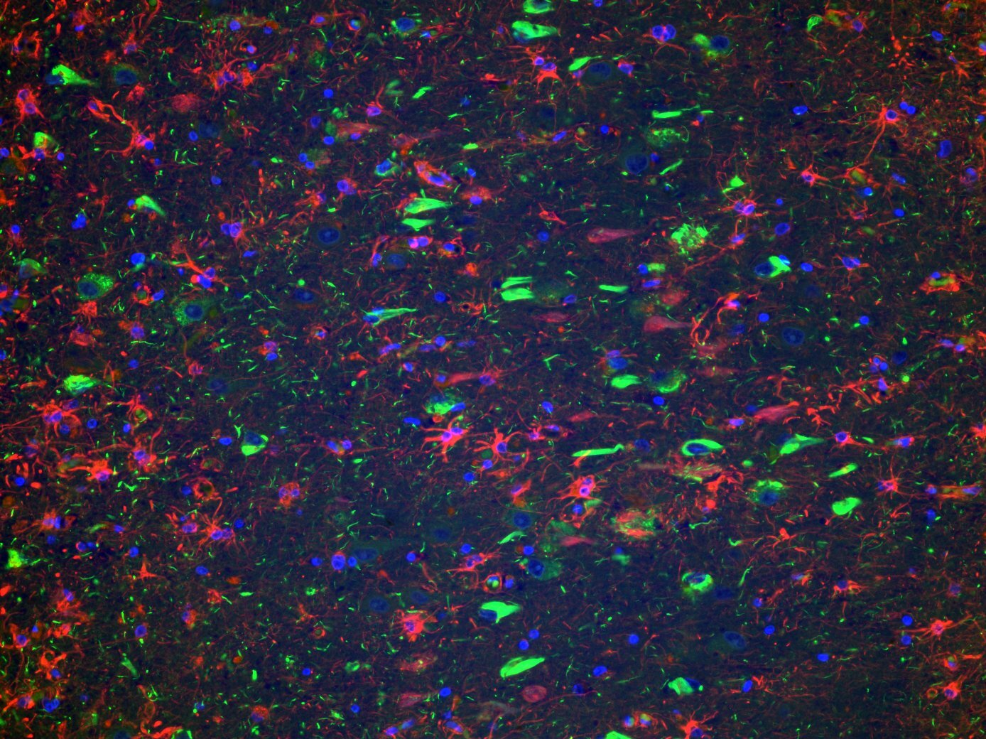
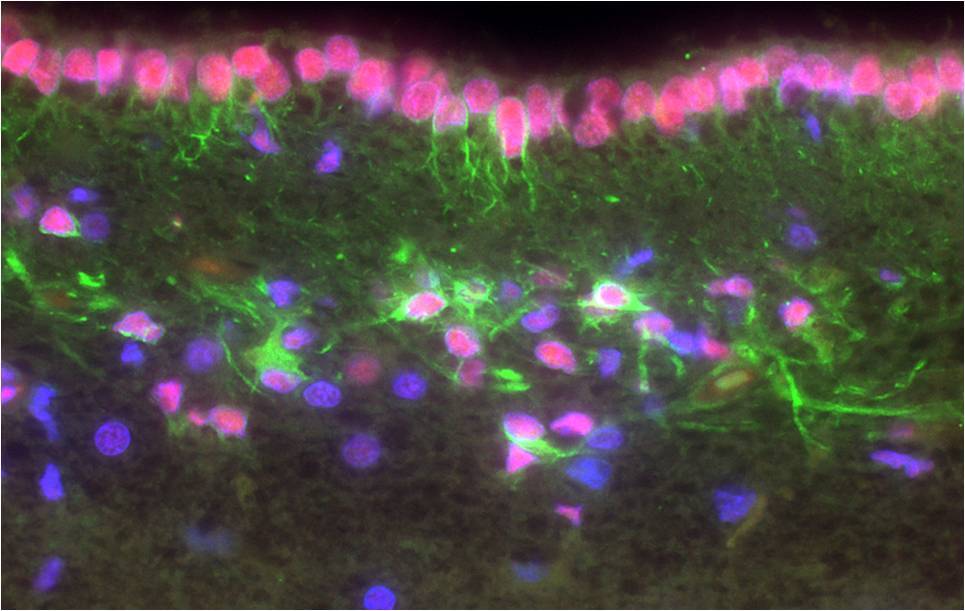
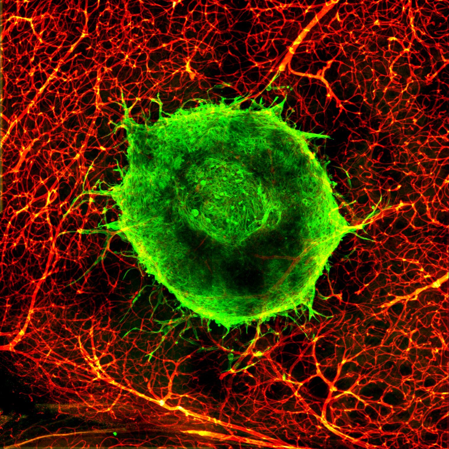
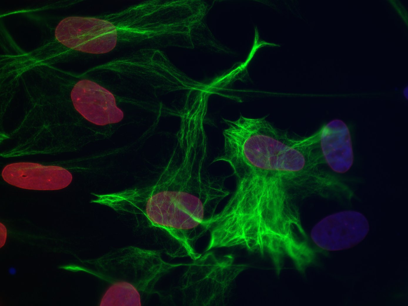
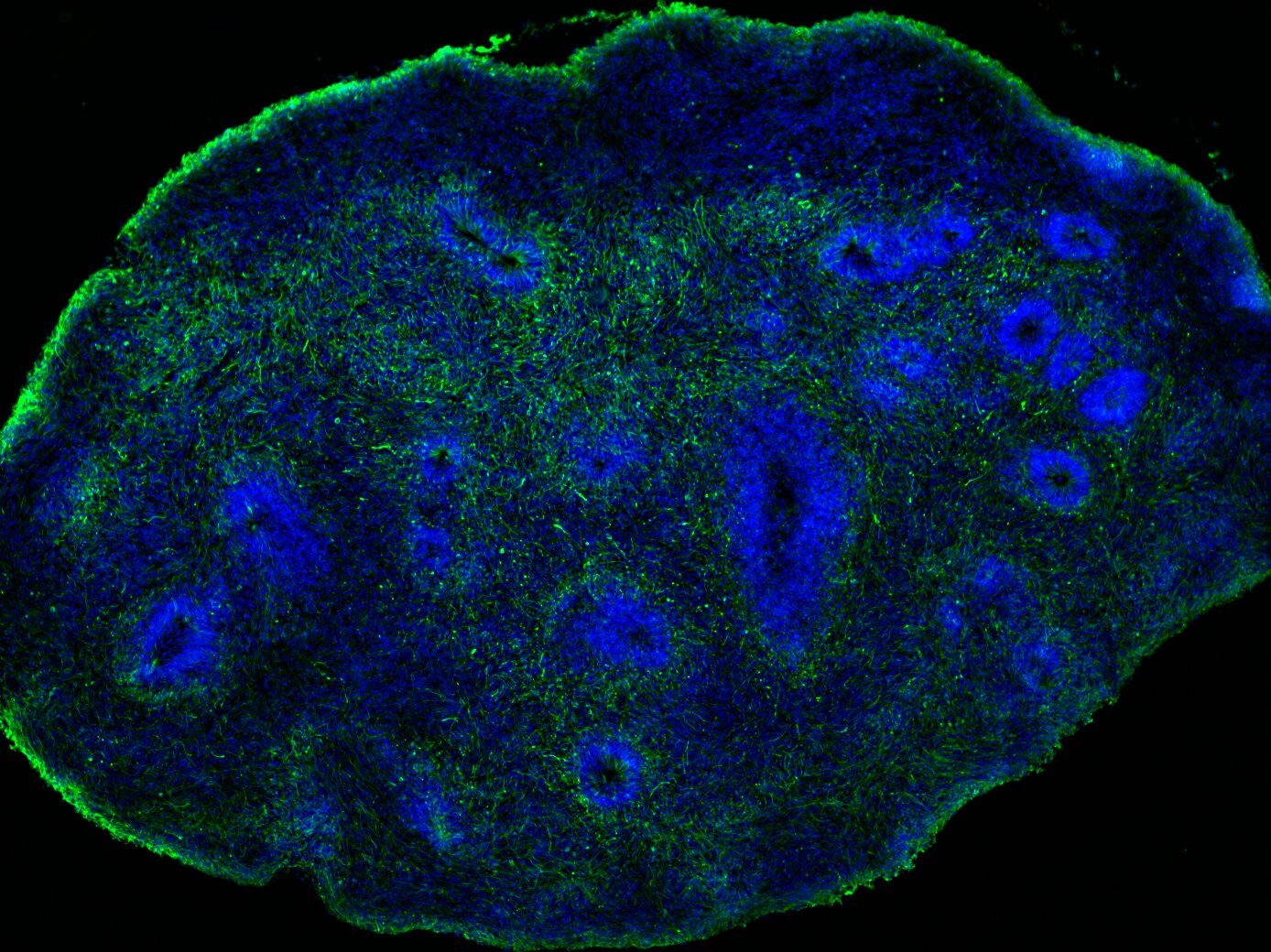
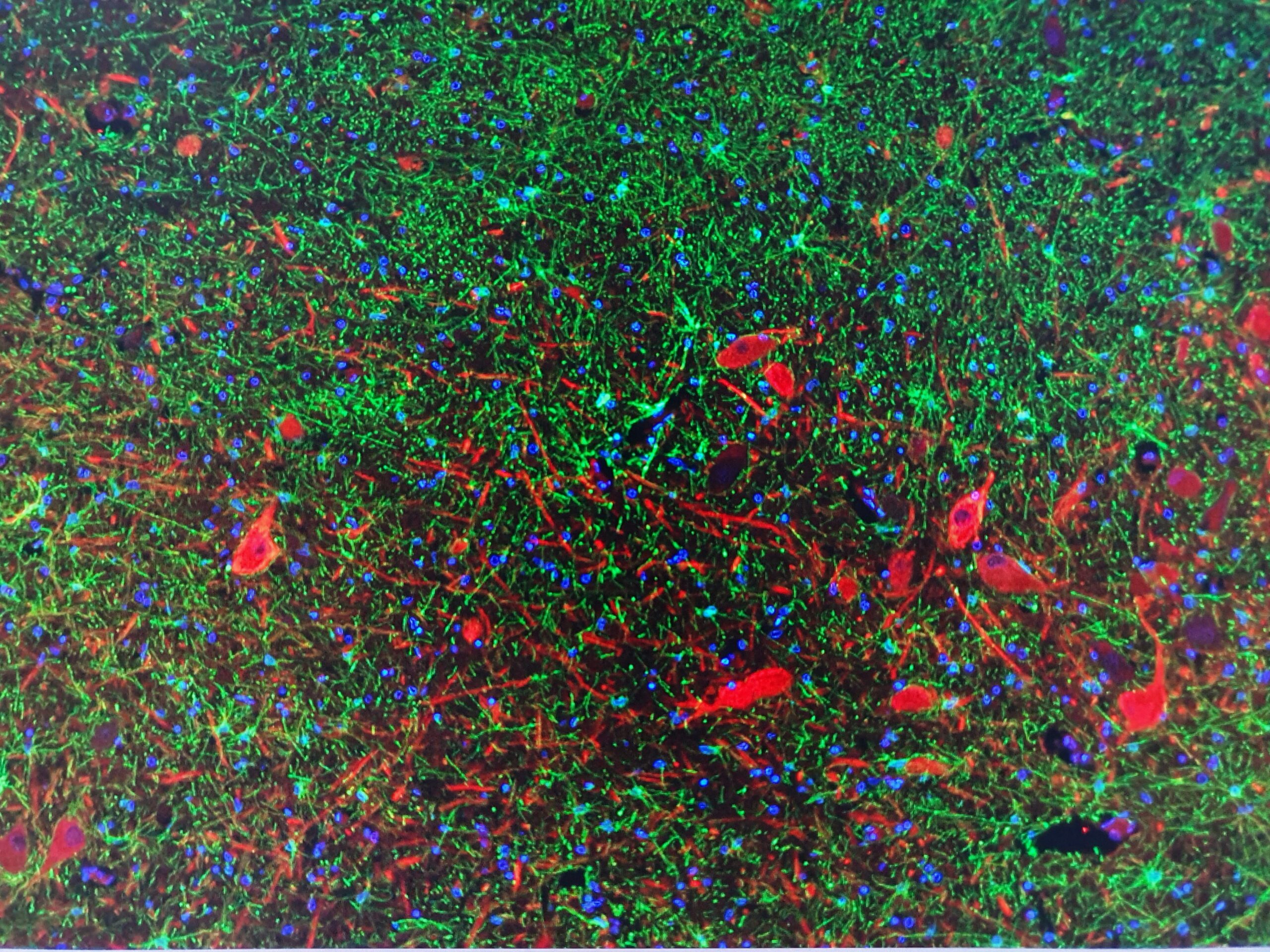
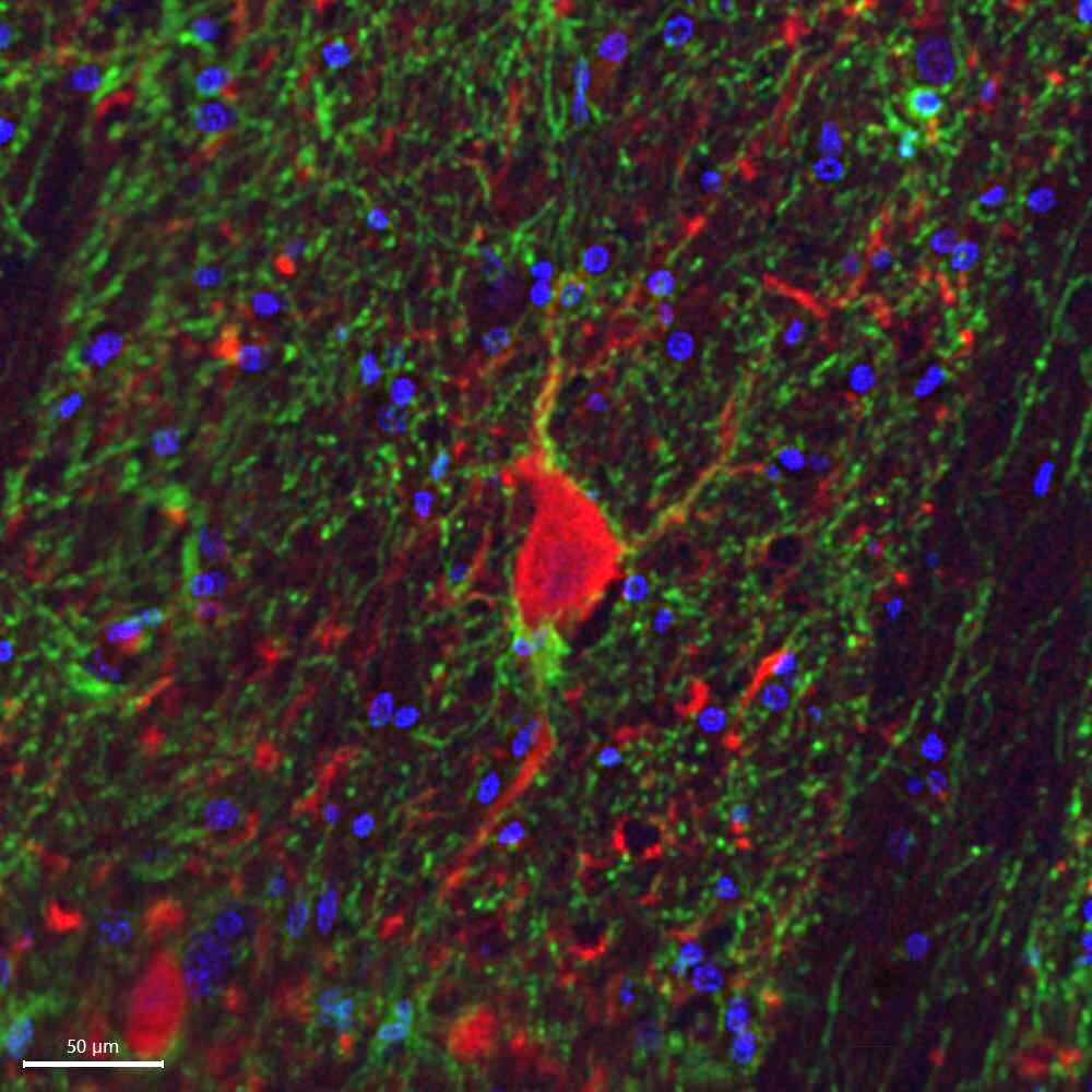
I am a glia biologist, and GFAP is my favourite protein. I am intrigued by the intermediate filament cytoskeleton and its role in human brain development, reactive gliosis, and glioma


Group members

Jacqueline Sluijs – Senior Research Technician
Lab coordinator and assistance with molecular- and cell biological experiments

Cell culture, viral production, human postmortem brain tissue, MACSorting, molecular cloning, PCR and Q-PCR, immunohistochemistry, genotyping, Bio-IP, westernblot


Danny van Nuijs – Research assistant
Helping around in the lab with data analysis and other lab work.

Genotyping, data analysis, microscopy


Charlotte Daemen – Research assistant
Studying astrocytes in an Alzheimer Mouse model with and without suppressed astrocyte reactivity.

Immunohistochemistry, cryostat, genotyping, confocal microscopy, mouse handling in a Novel Object Recognition and Barnes Maze task


Marloes Verkerke – PhD candidate
My research focusses on the effect of APOE4, the largest genetic risk factor for late onset AD, on neuron-glia interaction in iPSC models.

2D human iPSC models, human brain organoids, glia, calcium imaging


Werner Dykstra – PhD candidate
Modeling Alexander Disease with cerebral organoids

iPSC culture, generation and analysis of cerebral organoids


Anna van Regteren Altena – PhD candidate
My research focusses on the cellular and functional mapping of the human Ventral Tegmental Area in order to provide a single cell reference atlas to understand the involvement of cell types in reward system-related brain disorders.

Nuclei isolation, RNA sequencing, single cell-omics, 2D and 3D immunohistochemistry, human post-mortem brain tissue


Lois Kistemaker – PhD candidate
My research focuses on creating a human in vitro model that connects the blood-brain barrier to human brain organoids in order to test how therapeutic compounds enter and affect the (diseased) brain.
cerebral organoids, organ-on-a-chip

Lianne Hulshof – PhD candidate
Examining the role of astrocytes in Alzheimer’s disease by making use of a mouse model. Determining the differences between Alzheimer’s disease astrocytes and healthy controls, and testing whether changes in astrocyte gene expression can rescue memory deficits as observed in our mouse model.

Immunohistochemistry, MACSorting, sequencing, mouse breeding, memory tasks


Christiaan Huffels – Postdoc
Unravelling alterations in synaptic plasticity and neuron-glia interactions in early-stage Alzheimer’s disease.

Slice electrophysiology, calcium imaging, immunohistochemistry


Emma van Bodegraven – Assistant professor
My research aims to identify factors in the environment of brain tumors (glioma) which drive tumor invasion using 3D human brain tumor models. I focus on factors which shape the mechanical properties of the tumor environment and guide tumor invasion through mechanotransduction.

Primary glioma (2D/3D) cell culture, human iPSC-derived brain tumor models, single cell RNA sequencing and 3D invasion assay


Recent papers

  1. Huffels CFM, Middeldorp J, Hol EM. Aß Pathology and Neuron-Glia Interactions: A Synaptocentric View. Neurochem Res. 2022 Aug 17. doi: 10.1007/s11064-022-03699-6.Epub ahead of print. PMID: 35976488.
  2. Hulshof LA, van Nuijs D, Hol EM, Middeldorp J. The Role of Astrocytes in Synapse Loss in Alzheimer’s Disease: A Systematic Review. Front Cell Neurosci. 2022 Jun 16;16:899251. doi: 110.3389/fncel.2022.899251.PMID: 35783099; PMCID: PMC9244621.
  3. Hol EM, Pasterkamp RJ. Microglial transcriptomics meets genetics: new disease leads. Nat Rev Neurol. 2022 Apr;18(4):191-192. doi: 10.1038/s41582-022-00633-w.Erratum in: Nat Rev Neurol. 2022 Mar 8;: PMID: 35228701.
  4. van Asperen JV, Robe PAJT, Hol EM. GFAP Alternative Splicing and the Relevance for Disease – A Focus on Diffuse Gliomas. ASN Neuro. 2022 Jan-Dec;14:17590914221102065. doi: 10.1177/17590914221102065.PMID: 35673702; PMCID: PMC9185002.
  5. Hulshof LA, Frajmund LA, van Nuijs D, van der Heijden DCN, Middeldorp J, Hol EM. Both male and female APPswe/PSEN1dE9 mice are impaired in spatial memory and cognitive flexibility at 9 months of age. Neurobiol Aging. 2022 May;113:28-38. doi: 10.1016/j.neurobiolaging.2021.12.009. Epub 2022 Feb 12. PMID: 35294867.
  6. Huffels CFM, Osborn LM, Cappaert NLM, Hol EM. Calcium signaling in individual APP/PS1 mouse dentate gyrus astrocytes increases ex vivo with Aβ pathology and age without affecting astrocyte network activity. J Neurosci Res. 2022 Jun;100(6):1281-1295. doi: 10.1002/jnr.25042. Epub 2022 Mar 16. PMID: 35293016; PMCID: PMC9314019.
  7. Donega V, van der Geest AT, Sluijs JA, van Dijk RE, Wang CC, Basak O, Pasterkamp RJ, Hol EM. Single-cell profiling of human subventricular zone progenitors identifies SFRP1 as a target to re-activate progenitors. Nat Commun. 2022 Feb 24;13(1):1036. doi: 10.1038/s41467-022-28626-9. PMID: 35210419; PMCID: PMC8873234.
  8. van Bodegraven EJ, Sluijs JA, Tan AK, Robe PAJT, Hol EM. New GFAP splice isoform (GFAPµ) differentially expressed in glioma translates into 21 kDa N-terminal GFAP protein. FASEB J. 2021 Mar;35(3):e21389. doi: 10.1096/fj.202001767R. PMID: 33583081.
  9. Smit T, Deshayes NAC, Borchelt DR, Kamphuis W, Middeldorp J, Hol EM. Reactive astrocytes as treatment targets in Alzheimer’s disease-Systematic review of studies using the APPswePS1dE9 mouse model. Glia. 2021 Feb 25. doi: 10.1002/glia.23981. Epub ahead of print. PMID: 33634529.
  10. Escartin C, Galea E, Lakatos A, O’Callaghan JP, Petzold GC, Serrano-Pozo A, Steinhäuser C, Volterra A, Carmignoto G, Agarwal A, Allen NJ, Araque A, Barbeito L, Barzilai A, Bergles DE, Bonvento G, Butt AM, Chen WT, Cohen-Salmon M, Cunningham C, Deneen B, De Strooper B, Díaz-Castro B, Farina C, Freeman M, Gallo V, Goldman JE, Goldman SA, Götz M, Gutiérrez A, Haydon PG, Heiland DH, Hol EM, Holt MG, Iino M, Kastanenka KV, Kettenmann H, Khakh BS, Koizumi S, Lee CJ, Liddelow SA, MacVicar BA, Magistretti P, Messing A, Mishra A, Molofsky AV, Murai KK, Norris CM, Okada S, Oliet SHR, Oliveira JF, Panatier A, Parpura V, Pekna M, Pekny M, Pellerin L, Perea G, Pérez-Nievas BG, Pfrieger FW, Poskanzer KE, Quintana FJ, Ransohoff RM, Riquelme-Perez M, Robel S, Rose CR, Rothstein JD, Rouach N, Rowitch DH, Semyanov A, Sirko S, Sontheimer H, Swanson RA, Vitorica J, Wanner IB, Wood LB, Wu J, Zheng B, Zimmer ER, Zorec R, Sofroniew MV, Verkhratsky A. Reactive astrocyte nomenclature, definitions, and future directions. Nat Neurosci. 2021 Mar;24(3):312-325. doi: 10.1038/s41593-020-00783-4. Epub 2021 Feb 15. PMID: 33589835; PMCID: PMC8007081.
  11. Verkerke M, Hol EM, Middeldorp J. Physiological and Pathological Ageing of Astrocytes in the Human Brain. Neurochem Res. 2021 Feb 8. doi: 10.1007/s11064-021-03256-7. Epub ahead of print. PMID: 33559106.
  12. Snijders GJLJ, van Zuiden W, Sneeboer MAM, Berdenis van Berlekom A, van der Geest AT, Schnieder T, MacIntyre DJ, Hol EM, Kahn RS, de Witte LD. A loss of mature microglial markers without immune activation in schizophrenia. Glia. 2021 May;69(5):1251-1267. doi: 10.1002/glia.23962. Epub 2021 Jan 7. PMID: 33410555; PMCID: PMC7986895.
  13. Alsema AM, Jiang Q, Kracht L, Gerrits E, Dubbelaar ML, Miedema A, Brouwer N, Hol EM, Middeldorp J, van Dijk R, Woodbury M, Wachter A, Xi S, Möller T, Biber KP, Kooistra SM, Boddeke EWGM, Eggen BJL. Profiling Microglia From Alzheimer’s Disease Donors and Non-demented Elderly in Acute Human Postmortem Cortical Tissue. Front Mol Neurosci. 2020 Oct 28;13:134. doi: 10.3389/fnmol.2020.00134. PMID: 33192286; PMCID: PMC7655794.
  14. Antonovaite N, Hulshof LA, Hol EM, Wadman WJ, Iannuzzi D. Viscoelastic mapping of mouse brain tissue: Relation to structure and age. J Mech Behav Biomed Mater. 2021 Jan;113:104159. doi: 10.1016/j.jmbbm.2020.104159. Epub 2020 Oct 28. PMID: 33137655.
  15. de Sonnaville SFAM, van Strien ME, Middeldorp J, Sluijs JA, van den Berge SA, Moeton M, Donega V, van Berkel A, Deering T, De Filippis L, Vescovi AL, Aronica E, Glass R, van de Berg WDJ, Swaab DF, Robe PA, Hol EM. The adult human subventricular zone: partial ependymal coverage and proliferative capacity of cerebrospinal fluid. Brain Commun. 2020 Oct 13;2(2):fcaa150. doi: 10.1093/braincomms/fcaa150. PMID: 33376983; PMCID: PMC7750937.
  16. Snijders GJLJ, Sneeboer MAM, Fernández-Andreu A, Udine E; Psychiatric donor program of the Netherlands Brain Bank (NBB-Psy), Boks MP, Ormel PR, van Berlekom AB, van Mierlo HC, Bӧttcher C, Priller J, Raj T, Hol EM, Kahn RS, de Witte LD. Distinct non-inflammatory signature of microglia in post-mortem brain tissue of patients with major depressive disorder. Mol Psychiatry. 2020 Oct 7. doi: 10.1038/s41380-020-00896-z. Epub ahead of print. PMID: 33028963.
  17. Fernandez-Klett F, Brandt L, Fernández-Zapata C, Abuelnor B, Middeldorp J, Sluijs JA, Curtis M, Faull R, Harris LW, Bahn S, Hol EM, Priller J. Denser brain capillary network with preserved pericytes in Alzheimer’s disease. Brain Pathol. 2020 Nov;30(6):1071-1086. doi: 10.1111/bpa.12897. Epub 2020 Sep 29. PMID: 32876357.
WP Twitter Auto Publish Powered By : XYZScripts.com