GliaNed meeting 2022
Big success with the annual GliaNed meeting! The main goals of this informal meeting are to stimulate interaction between glia researchers in The Netherlands and to exchange knowledge. Elly Hol (translational neuroscience, UMCU) and Wia Baron (UMCG) put together a great program and were thrilled to have Prof. Inge Huitinga to open the show with her keynote lecture on ‘The role of microglia in MS and major depression’.
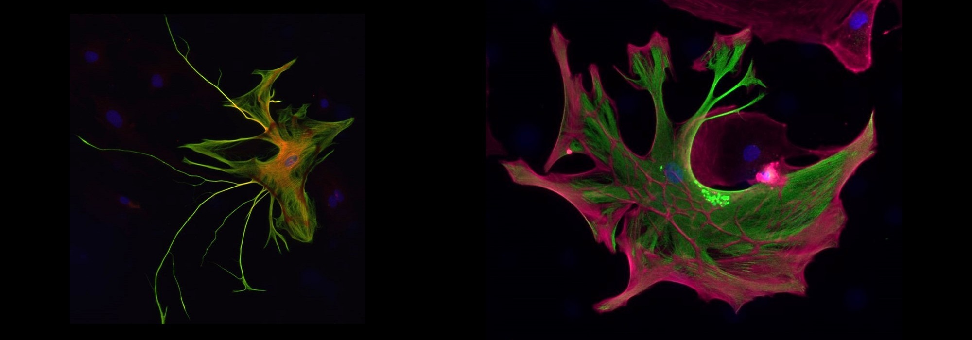
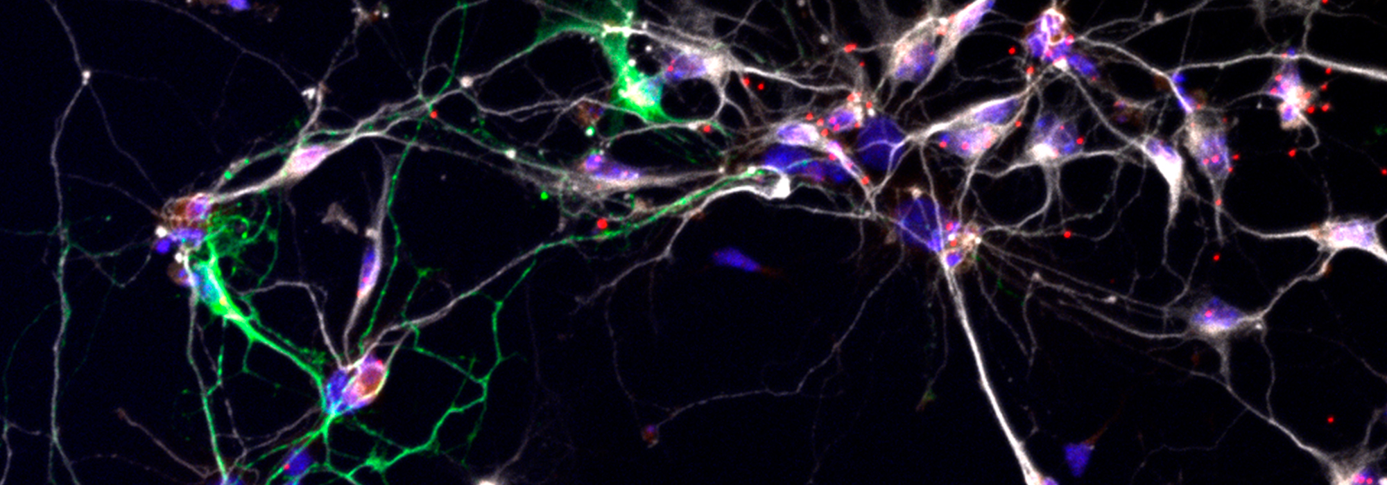
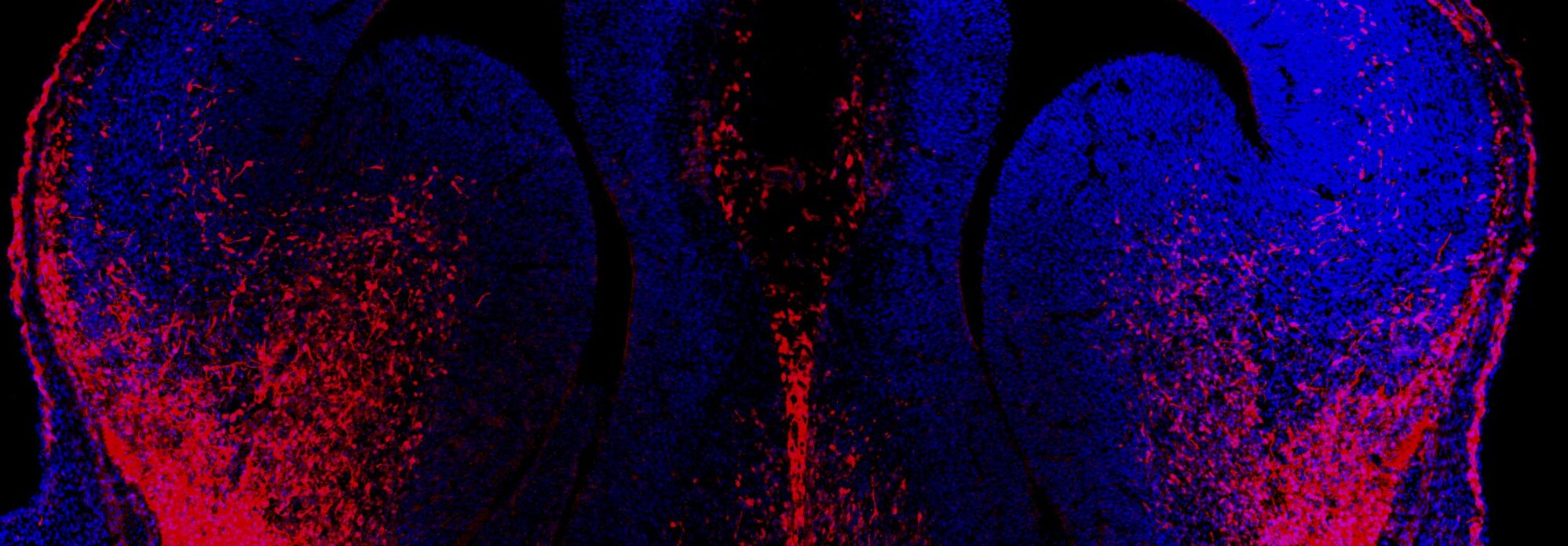
The meeting got additional contribution from our department. Carla Gomes da Silva presented her work on how oligodendrocyte precursors regulate cortical networks during mammalian development. Werner Dykstra explained how he studies the role of the astrocyte cytoskeletion in brain organoids. Several other researchers throughout the Netherlands have presented their latest research.

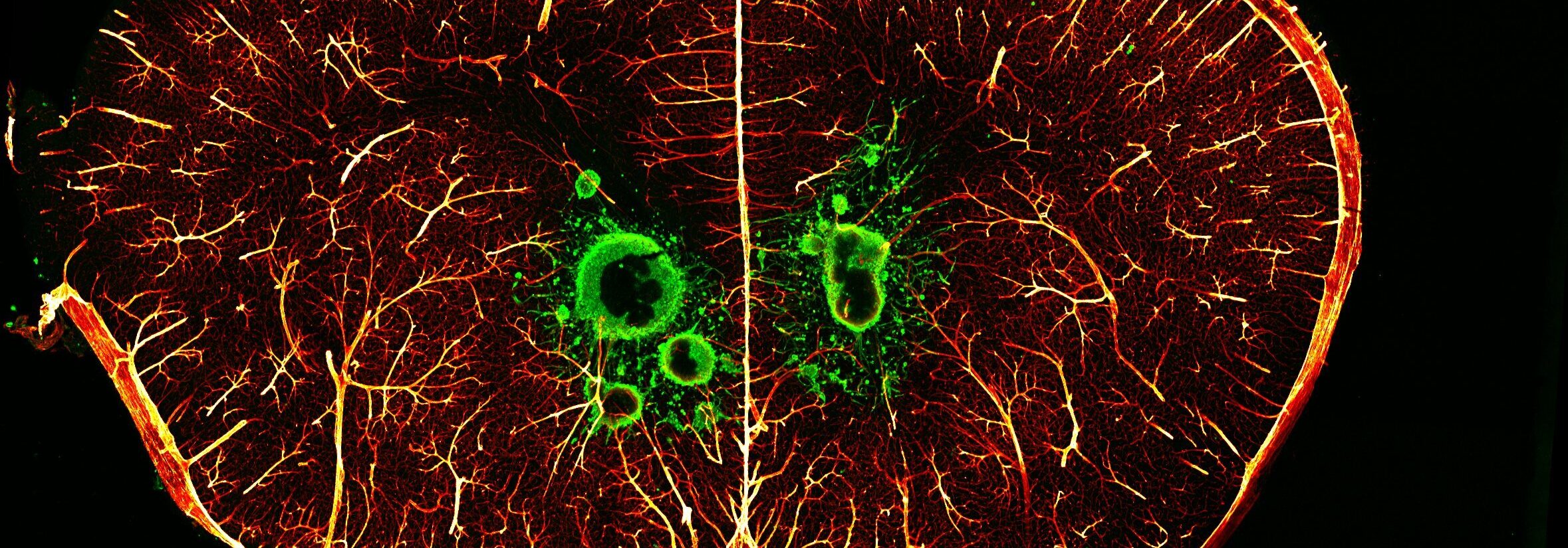
“It is very impressive how glia is involved practically in every brain process and disease you can imagine. GliaNed is providing an essential platform to bring researchers of this rapidly growing field together” Onur Basak
We hope to see the community at the next GliaNed meeting in Groningen.

 We proudly celebrate the inauguration of prof. Elly Hol of our