ENW-M-2 grant for Heidi Lesscher and Frank Meye on risky play and stress resilience

New ENW-M-2 grant for our researchers! Recently the NWO Domain Board Science approved twenty-six grant applications in the Open Competition Domain Science-M programme. One of these grants was awarded to Heidi Lesscher (PHS, DWM) and Frank Meye (Translational Neuroscience, UMC Utrecht) to study if early life play opportunity, and in particular ‘risky’ play, promotes the development of cognitive control and stress resilience in later life. In this project, rats experiencing different degrees of play during early life will be compared for their degree of cognitive control under stress. Expectations are that enhanced opportunities for risky social play in early life enhance stress resilience in later life. We will also address the neurobiological effects of social play on cognitive control networks. Understanding how play shapes stress resilience is important for the prevention of mental health problems. Stay tuned for positions opening up related to this project.
“Very excited to team up with Heidi Lesscher to unravel if early life risky play opportunities alter neural networks of cognitive control to confer stress resilience in later life” Frank Meye
This research will in part be carried out at our Translational Neuroscience Department, as part of the UMC Utrecht Brain Center.
Leptin targets spatially diverse neurons


Leptin is a hormone that is secreted by fat and signals the need to stop eating and increase energy expenditure via leptin receptors (LepR). Various hunger and reward centers in the brain contain different LepR expressing neurons. The primary leptin center is the well-studied arcuate nucleus. Other hypothalamic nuclei are less abundant in leptin receptor but also essential in encoding leptin’s actions. The composition of these LepR neurons have not been well understood.
In their recently published paper in Science Reports, Nefeli Kakava and colleagues from the UMC Utrecht Brain Center explore the scarce LepR population in the lateral hypothalamus. This population is known for its effects on food intake and food reward, and may be defective in eating disorders. The authors successfully capture the transcriptome of these neurons using TRAP-Seq. Exploration of their molecular profile confirms the expression of diverse neuropeptides and receptors. Microscopy analysis reveal their diverse spatial expression patterns. Moreover, they unravel new markers that could have significant role in energy balance. They also explore what is the transcriptional response of these neurons to energy deficit.
“I am excited that we have successfully managed to capture RNA from this very rare albeit significant population of leptin responsive cells and hope our findings inspire new research”
Nefeli obtained her PhD in 2020 as part of the Adan lab, where she developed viral vector tools to target and manipulate the activity of brain cells involved in food reward and energy balance. In collaboration with the Basak lab, she has profiled the hypothalamic LepR cells using TRAP-Seq and single cell genomics techniques.

Characterising social stress responsive ventral tegmental area neurons

In this study published at Frontiers in Behavioral Neuroscience, Ioannis Koutlas and colleagues of the Meye lab, use the expression of immediate-early genes to characterize social-stress activated neuronal subsets in the ventral tegmental area. They show that cells of different molecular identities (dopaminergic, GABAergic, glutamatergic and combinatorial neurons) that are dispersed throughout the entire VTA are activated by a social stress episode. Furthermore, they validate the use of targeted recombination in active populations (TRAP2) to capture this VTA stress-activated neuronal ensemble and make it tractable for further manipulations. Finally, the use of TRAP2 allowed them to look into intrinsic electrophysiological properties of these neurons and show that stress activated VTA cells are more excitable than neighbouring cells that were not activated by stress.

“I am very excited that this work is published. It gives insight on stress-encoding VTA neuronal populations and provides us with the tools to answer further exciting questions” – Ioannis
Cellular diversity of the developing mouse Habenula
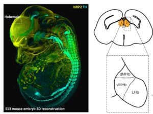
The habenula (Hb) plays a key role in processing reward information and mediating aversive responses to negative stimuli. In the recent issue of Cell Reports, Lieke van de Haar and colleagues have revealed how the cellular diversity of the mouse habenula is formed during development. In the work title “Cellular diversity of developing habenula may illuminate risk for human psychiatric disorders”, Lieke used single cell RNA sequencing, a technique that allows quantification of gene expression in thousands of cells simultaneously.
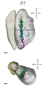 Reconstruction of lineage trajectories using computational tools revealed paths to habenula cell types. Intersectional genetics experiments by Oxana Garritsen showed that these include a physiologically distinct neuronal cell type that innervate the dorsal IPN which can be identified by Cartpt expression. Work by Lieke and Youri Adolfs using iDISCO and light sheet microscopy have shown that these cells localize to the medial habenula. The ePhys work by Danai Riga of the Meye lab found that these Cartpt+ neurons are electrophysiologically different from surrounding neurons. Finally, MAGMA analysis by Juliska Boer, who did her bachelor internship under the supervision of Lieke, revealed that developing cell populations align with human genetic risk loci of psychiatric disorders, such as the major depressive disorder.
“The habenula plays an important role in negative experiences, and is located dorsally to the thalamus and ventrally to the hippocampus…. we were interested in how the cells of the habenula are born and mature.” Lieke
Lieke performed her PhD work at the Pasterkamp lab of our Translational neuroscience department of the UMC Utrecht Brain Center. This work is a fine example of internal collaborations in the department which connect molecular, computational, genetic and electrophysiological techniques.
New Scientist Live! Hersenziekten in Tivoli
On Tuesday the 17th of May UMC Utrecht Brain Center and New Scientist organized ‘NewScientist Live! Hersenziekten in TivoliVredenburg. Several colleagues focused on the technologies of the future. It was an interesting evening, filled with exciting and necessary advances in brain research and patient care.

Foto: Bob Bronshoff
Foto: Bob Bronshoff
Prof. dr. Jeroen Pasterkamp and PhD student Tiziana Hey, from our own Translational Neuroscience department, starred on stage. Jeroen Pasterkamp started the evening off, highlighting important microscopic and organoid research performed in his lab. Tiziana Hey, together with 2 fellow young scientists, closed the evening, explaining research with the use of single cell sequencing techniques. Interested in reading more about this informative evening? You can find a summary of the evening on the website of New Scientist or watch the aftermovie (in Dutch).
“Brain research is like a black box: one big fascinating puzzle” T. Hey
We are very glad to disseminate the knowledge generated at the UMC Utrecth Brain Center.